Note: Your progress in watching these videos WILL NOT be tracked. These training videos are the same videos you will experience when you take the full ProPALS Recertification program. You may begin the training for free at any time to start officially tracking your progress toward your certificate of completion.
Sinus tachycardia is a common response to a variety of conditions. It's often associated with a child who is:
- Anxious
- Crying
- Febrile (has a fever)
- Ill or injured
In this lesson, we'll look more closely at an example of what sinus tachycardia looks like on an ECG for a pediatric patient and see what findings and measurements lead us to that conclusion.
Pro Tip #1: When treating a tachycardic child without signs and symptoms of cardiac compromise, you should search for the underlying cause of that patient's tachycardia.
For a tachycardic child with a pulse and not in cardiac compromise, you should assess for signs of:
- Hypotension
- Altered mental status
- Shock
- Other life-threatening hemodynamic instabilities
Pro Tip #2: What does hemodynamic instability mean? It means that unless healthcare providers do something, the process is unstable and in danger of failing. Some common examples of hemodynamic instability include circulatory collapse, shock (particularly decompensating shock), hypoperfusion, and cardiovascular failure.
If you discover signs or symptoms of any of those during your initial assessment, management includes supporting the airway, breathing, and circulation (or the ABCs) of the patient, which include:
- Providing oxygen as needed
- Obtaining vital signs, including blood pressure and pulse oximetry
- ECG monitoring or by attaching defibrillator pads
- Establishing IV/IO vascular access
Now let's take a look at an ECG for a patient in sinus tachycardia.
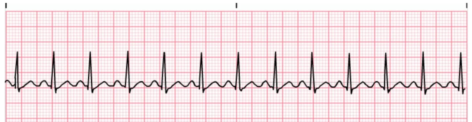
*Sinus Tachycardia ECG
1. The Heart Rhythm
The first thing you'll want to look at is the heart rhythm. Does the heart rhythm look regular? Or does it look irregular? In the above graphic, it's regular.
2. The Heart Rate
Next, you'll want to look at the heart rate of the patient. What is the patient's heart rate? Is it normal? Or is it too slow or too fast? In this case, it's too fast, as the rate is 120 beats per minute.
Remember, the definition of a normal heart rate will vary based on the child's age. For example, the normal heart rate for an 11-year-old patient might be bradycardic for an infant.
3. P-Wave
After looking at the heart rate, check to see if the patient's P-waves look normal by asking yourself the following few questions.
- Are the patient's P-waves present? In this case, the answer is yes.
- Do they occur regularly? The answer is yes again.
- Is there one P-wave for each QRS complex? Yes, there is.
- Are the P-waves smooth, rounded, and upright? The answer is again yes.
- Do all the P-waves have a similar shape? Yes, they all have a similar shape.
4. PR Interval
Next, look at the PR interval on the patient's ECG readout and ask yourself the following questions:
- Is the PR interval normal, meaning less than .20 seconds or is it contained within one large square on the readout? The answer is yes, it's less than .20 seconds and contained within one large square.
- Is the PR interval constant? Yes, it is.
5. QRS Complex
The last thing you should look at to determine if the sinus rhythm is normal or not is the QRS complex and ask yourself these questions while you do:
- Is the QRS interval less than .09 seconds? Yes, it is.
- Is the QRS complex wide or narrow? In this case, it's narrow.
- Are the QRS complexes similar in appearance or are there noticeable differences? In this case, we can see that each looks similar.
So, what is your cardiac interpretation? Based on these questions and on the findings from the ECG readout above, it's safe to say that this patient is in sinus tachycardia.
- We have a regular rhythm.
- We have a faster than normal heart rate, at 120 beats per minute.
- The P-waves look normal, with each being followed by a QRS complex.
- The PR interval is less than .20 seconds.
- The QRS is less than .09 seconds.
Unless you see signs of circulatory compromise in this patient, direct your attention to finding and treating the underlying cause for the tachycardia. But if the patient is unstable, rapid and effective treatment must be provided to correct the cause of the tachycardia.
Additional Sinus Tachycardia Information
Tachycardia can be a sign of a serious condition. A heart rate that is greater than 180 beats per minute in an Infant or toddler, and greater than 160 beats per minute in any child two years old or older, warrants further assessment.
Hypotension
Hypotension can be a threatening sign of imminent cardiac arrest in pediatric patients.
When hypotension develops in a pediatric patient who's in shock, physiologic compensatory mechanisms — like tachycardia and vasoconstriction – have likely failed. Hypotension with hemorrhage is thought to be consistent with an acute loss of 20 to 25 percent of circulating blood volume.
Hypotension in septic shock can occur from loss of intravascular volume and inappropriate vasodilation or severe vasoconstriction and inadequate cardiac
output.
The development of bradycardia in a child with hypotension and poor perfusion is a threatening sign. Management of the patient's airway and breathing and support of adequate intravascular volume, cardiac function, and perfusion are required to prevent cardiac arrest.
Hypoxemia
Hypoxemia occurs when your blood oxygen levels fall below a certain point. This can result in shortness of breath, headache, and confusion or restlessness.
As it pertains to this lesson, tachycardia may also develop in response to hypoxemia, as a means of increasing cardiac output. As tissue hypoxia worsens, these signs of cardiopulmonary distress become more severe.


